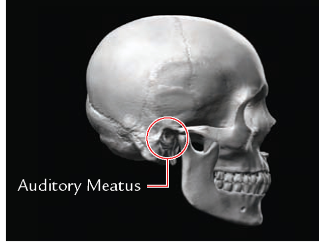

The temporal bones comprise the lateral skull base, forming portions of the middle and posterior fossae. It is helpful to examine the region in an organized and systematic fashion, going through the same checklist of key structures each time. There are a limited number of structures and disease entities in the temporal bone with which one must be familiar in order to proficiently interpret a computed tomographic (CT) or magnetic resonance (MR) imaging study of the temporal bone. For this journal-based CME activity, author disclosures are listed at the end of this article. The ACCME requires that the RSNA, as an accredited provider of CME, obtain signed disclosure statements from the authors, editors, and reviewers for this activity. Physicans should claim only the credit commensurate with the extent of their participation in the activity. The RSNA designates this journal-based activity for a maximum of 1.0 AMA PRA Category 1 Credit TM. The RSNA is accredited by the Accreditation Council for Continuing Medical Education (ACCME) to provide continuing medical education for physicians. Distinguish between various tumors of the temporal bone based on CT and MR findings.Describe imaging features of some common inflammatory conditions in the temporal bone.Recognize clinical presentations associated with important inflammatory and neoplastic conditions in the temporal bone region.Identify important anatomic landmarks in the temporal bone.After reading the article and taking the test, the reader will be able to:


 0 kommentar(er)
